
Equine athletes, much like their human counterparts, are prone to various musculoskeletal issues that can impede their performance and overall well-being.
Lameness and soreness are common afflictions among horses, often stemming from underlying injuries or strains.
Traditional methods of diagnosing such issues have limitations, making it challenging to pinpoint the exact source of discomfort.
However, with advancements in technology, equine healthcare has seen significant improvements, one of which is thermographic imaging.

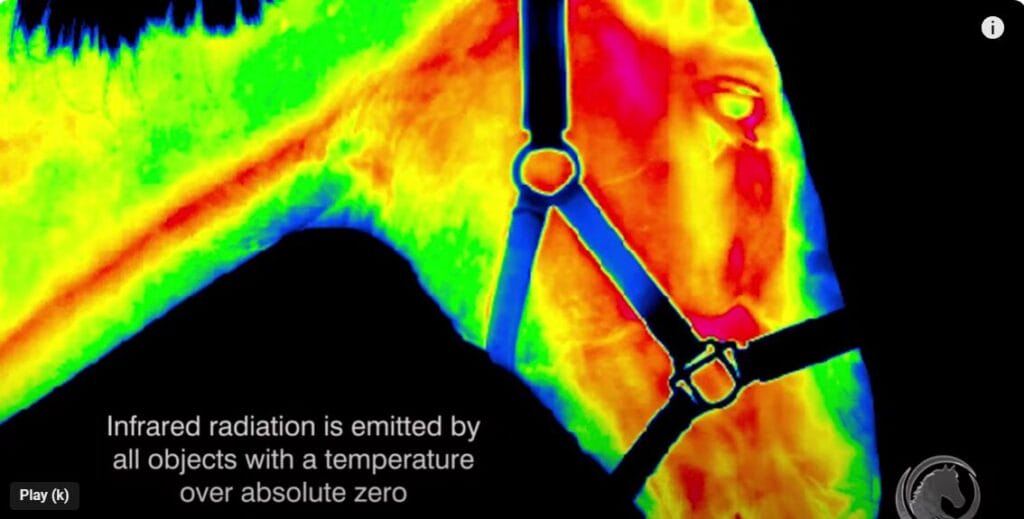
Thermographic imaging, also known as infrared thermography, is a non-invasive diagnostic tool that measures temperature variations on the surface of the skin.
In recent years, its application in equine medicine has gained traction, offering veterinarians and horse owners valuable insights into the physiological condition of their equine companions.
Understanding Thermographic Imaging
Thermographic imaging relies on infrared cameras to detect and visualize heat patterns emitted from the body. The principle behind this technology is simple: areas of inflammation or increased blood flow generate more heat, which is captured by the camera and displayed as colorful images.
By analyzing these thermal patterns, veterinarians can identify potential areas of concern, such as inflammation, muscle strain, or nerve damage, even before they manifest as clinical symptoms.